
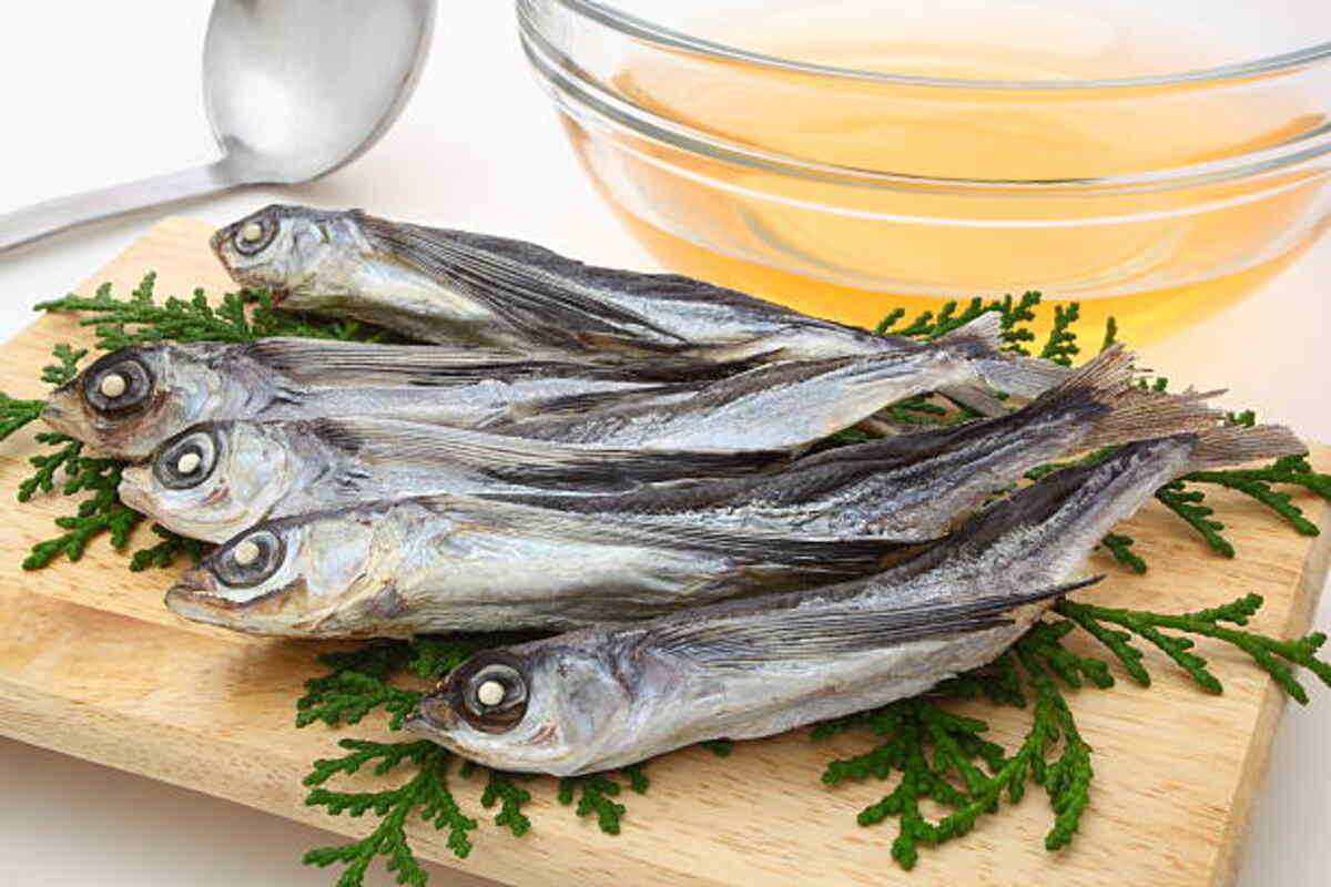
In this lab, you will study the fish skulls of bony fish species. Discover its parts and what each component does.
Begin by boiling a head from a bony fish (not recommended for cartilaginous species), allow it to cool, and pull away flesh while noting where bones come apart. Take notes as each bone disassembles.
Characteristics
Fish skulls are two-part structures composed of two encased bone “boxes.” Their exterior layer consists of dermal bone, while their interior, which surrounds the brain, is composed of endochondral bone. Furthermore, fish skulls include opercular bones that cover their gill surface and help bring oxygen into their system through breathing.
Fish are unique among vertebrates in that their skeleton is composed of both bones and cartilage, making it hard to determine the appearance of jawed vertebrates in their ancestral state, given that bony fish and cartilaginous fish skeletons differ dramatically from one another.
A fossil skull discovered in Estonia challenges this perception; Janusiscus was an ancient fish with a highly complex head and features that parallel those seen among other vertebrates.
A primary function of fish skulls is protecting their brains, with an outer shell called the neurocranium serving this function and being made from skeletal elements ossified in bony fishes or replaced by endochondral bone in chondrichthyans. Inside is the roof is, known as the splanchnocranium, containing branchial arches, which form part of their mouth mechanism or gill apparatus in teleosts and tetrapods, respectively.
Fish skulls do more than protect their brains; they can also serve as effective hiding places. Their outer surfaces are covered in thick scales that protect them from predators. This feature makes the fish worthwhile when hiding to escape danger or defend its territory.
A fish skull is an anchor for its tail fin, helping it remain upright and balanced in the water by using its tail fin to propel forward. Furthermore, dorsal fins help steer fish toward specific directions when swimming; these dorsal fins also maintain upright posture when resting.
Structure
A fish’s skull is an intricate structure of several small bones covered with cartilage that allow flexibility. The shape and composition of this structure are crucial to its survival; protecting its brain while permitting fast movements to escape larger predators or catch prey quickly are critical considerations for survival. Furthermore, each bone must be light enough for easy aquatic travel.
Fish skulls can be studied using various approaches, from micro-anatomy and ontogenetic development to computer simulations that explore their mechanics – these studies can shed light on how fish use their skulls for specific behaviors such as swallowing and biting.
Computer models can show how the bones of a fish’s head move during suction feeding when its muscles contract to expand its throat cavity and form a flow of water that draws in prey. Furthermore, this model shows how this flow is controlled by motions in different parts of its skull, such as its hyoid bone near the floor of its mouth cavity and the large operculum at the top of its head.
Another study utilized 3D morphometrics to compare Notothenioid species skulls using 3D morphometrics. Their results demonstrated a correlation between the size of fishes’ heads and their foraging niche; wider-skulled, shorter-jawed species tend to feed on bottom fish, while narrower-skulled, longer-jawed ones serve as scavengers and hunt for aquatic creatures in the water column. Furthermore, this research demonstrated that just 7 of 19 DoF (Dof) were sufficient to describe most movements involved during in-vivo suction feeding behavior of channel catfish skulls during in-vivo suction feeding behavior of this fish species.
Bony fishes’ skulls are more complex than lamprey or hagfishes’; however, even this complexity cannot compare to that of sharks, whose skull is known as the chondrocranium and comprised of neurocranium, splanchnocranium, and dermatocranium components.
Functions
A fish’s skeleton supports and protects its delicate organs while aiding bodily movement by providing levers for muscles to use bones as levers. Bony fishes have predominantly skeletal frameworks, while cartilage fishes possess more flexible frameworks made up mainly of cartilage (Fig 4.50).
The fish skeleton contains holes, hinges, and pockets that provide space for its internal organs. For instance, its brain is protected by its skull – comprised of small bones surrounding its core and an air intake hole at its front; additionally, these bones help form its head shape and create an opening for its mouth containing jaws and gill filaments.
Like other vertebrates, fish have an elaborate circulatory system to transport blood. Contraction of the heart muscle pumps blood into arteries with valves designed to ensure one-directional blood flow; blood low in oxygen but rich in carbon dioxide passes to the gill filaments where more oxygen is absorbed via capillaries before returning into their veins for circulation back through veins back into their bodies.
Fish have various senses, including lateral lines that detect vibration and movement in the water; compound eyes with multiple layers that can focus on objects at different distances; photophores, which provide hiding spaces or warn predators; as well as photophores used for hiding, warning predators or mating purposes. Some fish species produce light via chemical reactions in their bodies or from bacteria living on their skin that illuminate it at night or day – appearing luminescent during either nocturnal or daytime periods.
Fish have an intricate nervous system, though their brains may not be as significant. Bony fishes’ brains can be divided into sections for processing vision, learning, and motor responses while their forebrain controls smell; bony fish with well-developed scent sense tend to possess larger forebrains.
A fish’s digestive and excretory systems work in concert to bring food into their bodies, process it into usable nutrients for use by their cells, and discard any waste material or leftover food items through its mouth and teeth before being transported through its stomach, intestines, and anus (Fig 4.51); some waste is even expelled through its gills.
Evolution
Fishes exhibit exceptional control of fluid flow and movement into, within, and around their mouths for filtering, mouthbrooding, suction feeding, and prey processing. Fish achieve these feats without using hands or flexible muscular tongues but instead through the use of over 20 movable skeletal elements in their highly kinetic skulls – such as mandibular and hyoid arches, branchial apparatus supporting their gills, shoulder girdle, and in some species, even pharyngeal jaws (Gibb and Ferry-Graham 2005; Van Wassenbergh et al. 2016).
Long ago, it has remained unclear how jawless fish evolved into four-legged vertebrates, but new 3D fossils of Eriptychius Americans from Colorado’s Harding Sandstone formation may shed some light. The fossil shows an articulated neurocranium unlike anything found today among jawed hagfish or lampreys, with ten long cartilage pieces connected by scales without being fused, threading through canals to deliver blood or connect sensory organs.
Eriptychius fossils offer insight into how jawless fish skulls have developed into complex arrangements of jaws, bones, and movable segments found in living teleosts. Through experimental investigations of this fossil fish’s jawless skull development into modern-day living teleosts’ complex array of jaws, bones, and movable segments that support suction-feeding behavior, researchers are investigating how these movable parts generate the force required to capture prey during suction-feeding and have also assessed mechanical models of how this movement occurs before comparing these with observations of suction feeding behavior observed during life in real-time words of suction feeding behavior observed during suction feeding behavior observations in living teleosts.
Experimental tests corroborate previous computational simulations that determined that channel catfish skulls have 19 theoretical degrees of freedom (DoFs), yet most in vivo motion is limited to seven DoFs. The other DoFs seem related to how the skull expands depending on factors like the angle between an elliptical opening and curved canal, the speed of fluid flow inside the braincase, and soft tissues within its borders (Van Wassenbergh et al. 2015).